📄 Digital book in PDF · read it on your phone or print it
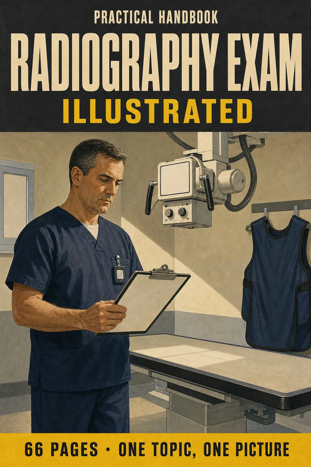
X-ray physics, mAs and kVp, the inverse square law, dose limits, grids, patient ID, contrast reactions and positioning with the central ray. Each topic gets its own page: the drawing first, then what you need to remember. 66 illustrated pages.
🔒 Secure checkout · PDF by email · 30-day guarantee
Every page has one big drawing and a box with the numbers, angles and steps you need to know:


Swipe to see more →
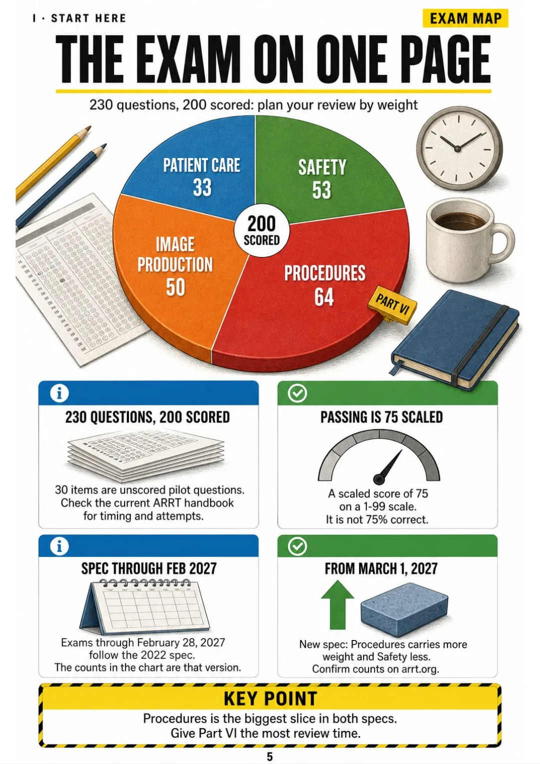
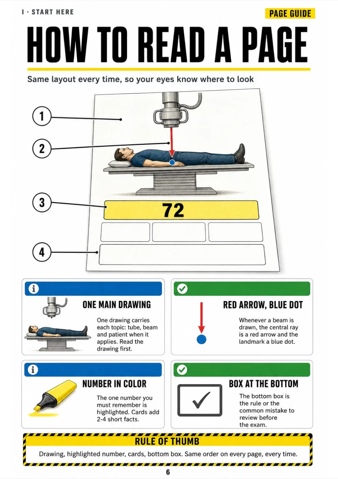
These are real pages from the book. The table of contents is the full one: 7 parts and 62 topics.
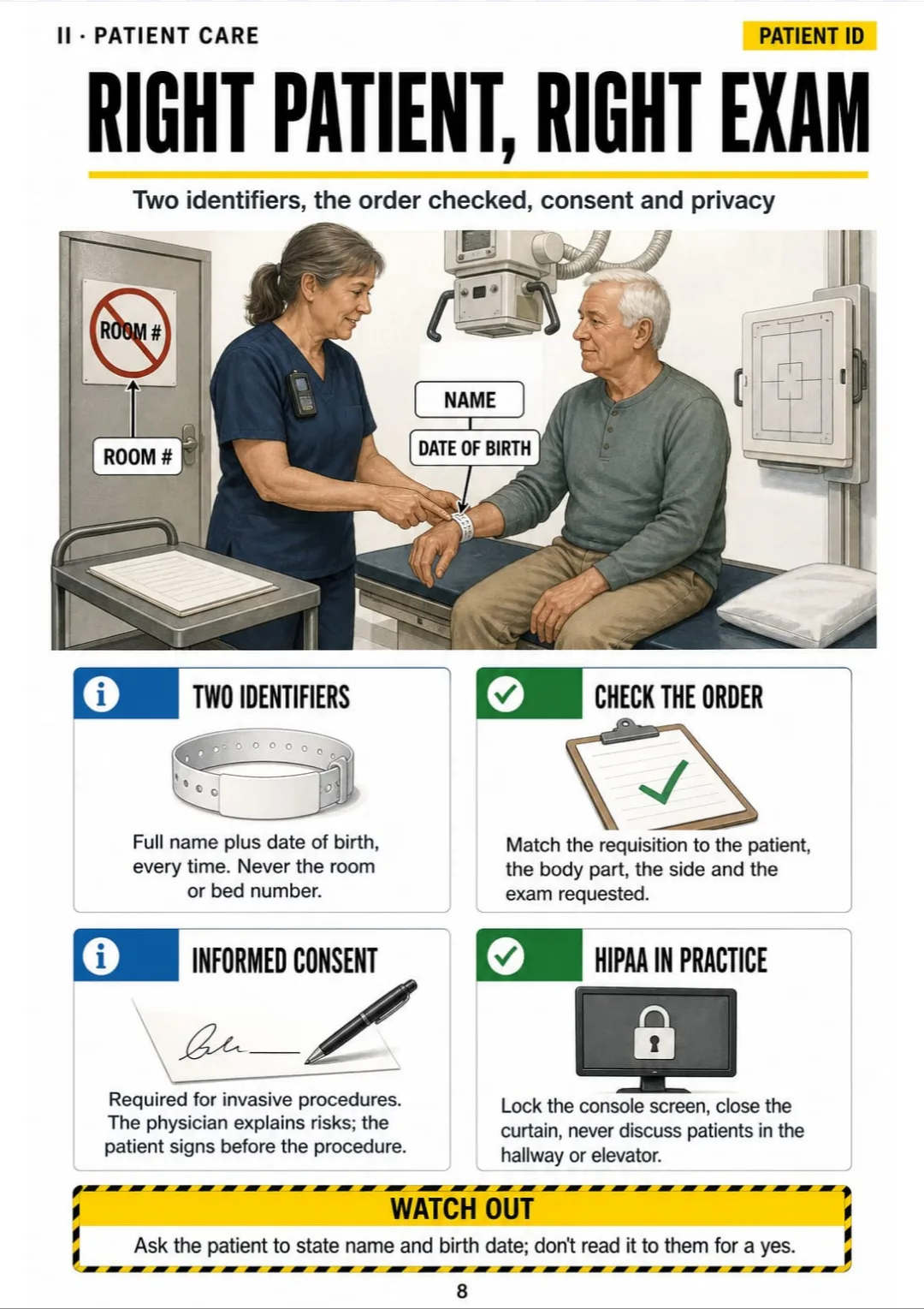
You're in clinicals with the requisition in your hand, and the tech nods toward the patient: "Go ahead, ID her." For a second you freeze. The wristband? The room number? What do you ask first?
Your textbook covers patient identification in three dense paragraphs, somewhere between consent, privacy and outpatient policy. Nothing on that page shows you what to actually do at the door.

In this book, the same topic is one page: the patient, the wristband and the requisition drawn side by side. Two identifiers every time, name and date of birth, never the room number, in a box you see at a glance.
The rest works the same way: the inverse square law, the 15% rule, grid cutoff, the PA chest with its central ray, contrast reactions. Each one has its own page.
So you understand it, and you do it in front of the tech without going back to the manual.

Start here, patient care, x-ray physics, radiation safety, image production, positioning and procedures, and a final review: 66 pages, all illustrated
Think of it this way:
You're in the room for an AP knee. The tech asks how you'll angle the tube, and you need the answer now, not after flipping through a whole chapter.
In the book you open the knee page and see the leg drawn from the front: the central ray as a red arrow, the angle right next to it, and the ASIS-to-tabletop measurement that decides it.

Same with mAs and kVp, the inverse square law, the four grid errors, the dose limits and the PA chest: each one on its own page.
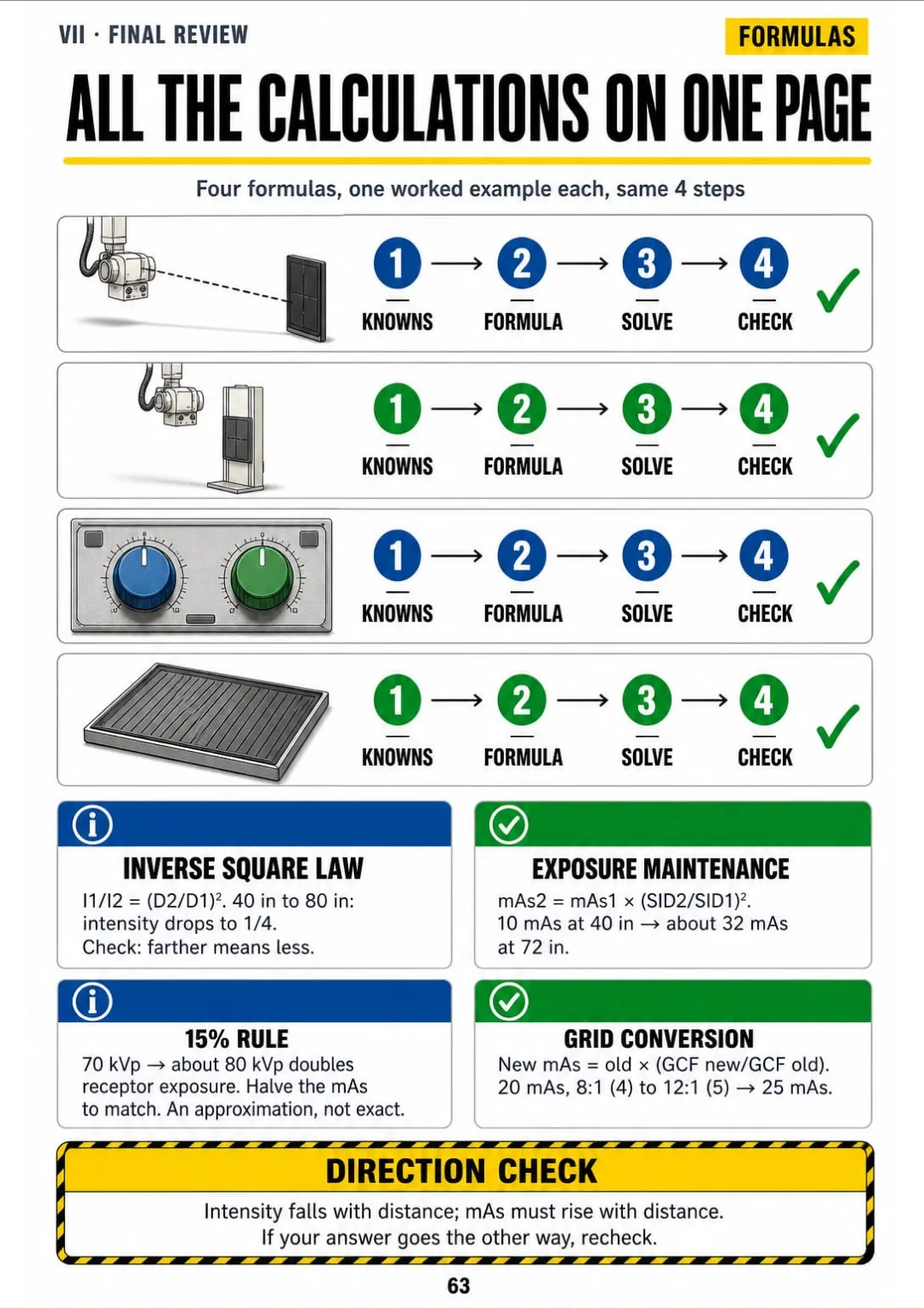
Radiography Exam Illustrated is 7 parts and 62 topics, all drawn, and at the end all the calculations on one page, the numbers to know cold and a week-before checklist.
What you see drawn, you don't have to memorize.
The exam on one page, how to read every page in this book and the 4 steps every calculation follows.
Patient ID and consent, vital signs, precautions, sterile technique, transfers, tubes and lines, contrast media and reactions, emergencies in the room.
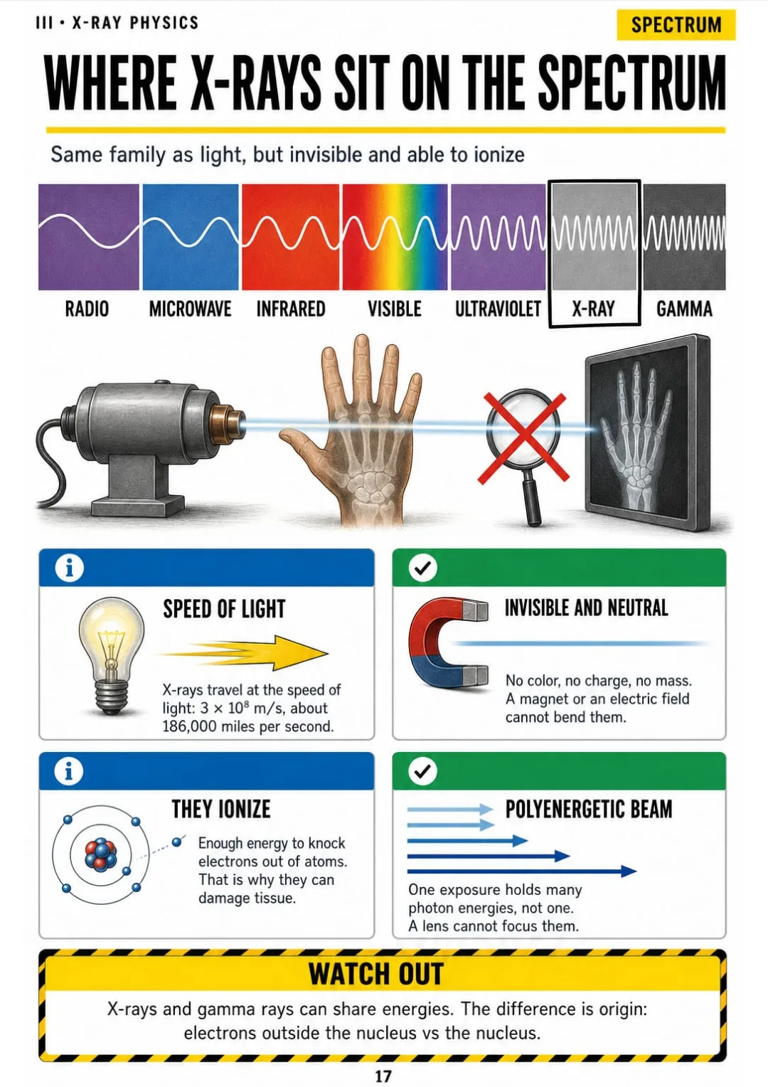
The spectrum, inside the tube, brems vs characteristic, focal spot, the heel effect, photon interactions, filtration, HVL and radiation units: the physics you've read ten times, as pictures you can follow.
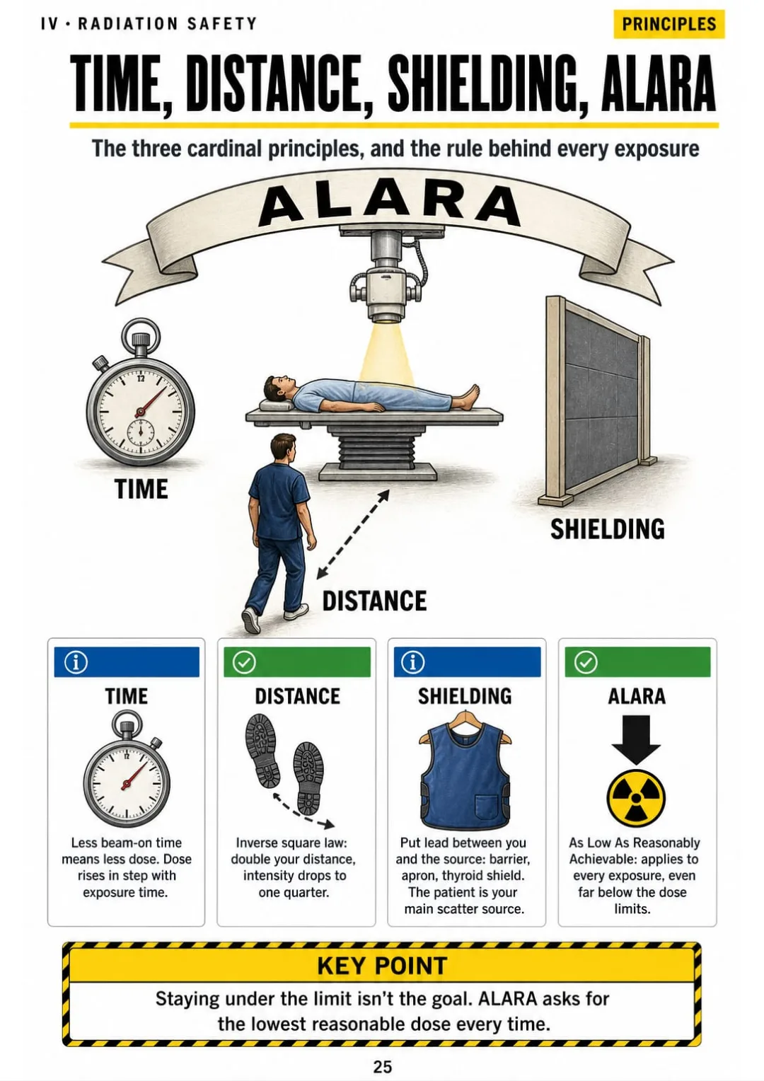
Time, distance, shielding and ALARA, dose limits, pregnancy, aprons and barriers, protecting the patient, radiation biology, dosimeters, fluoro and mobile safety.
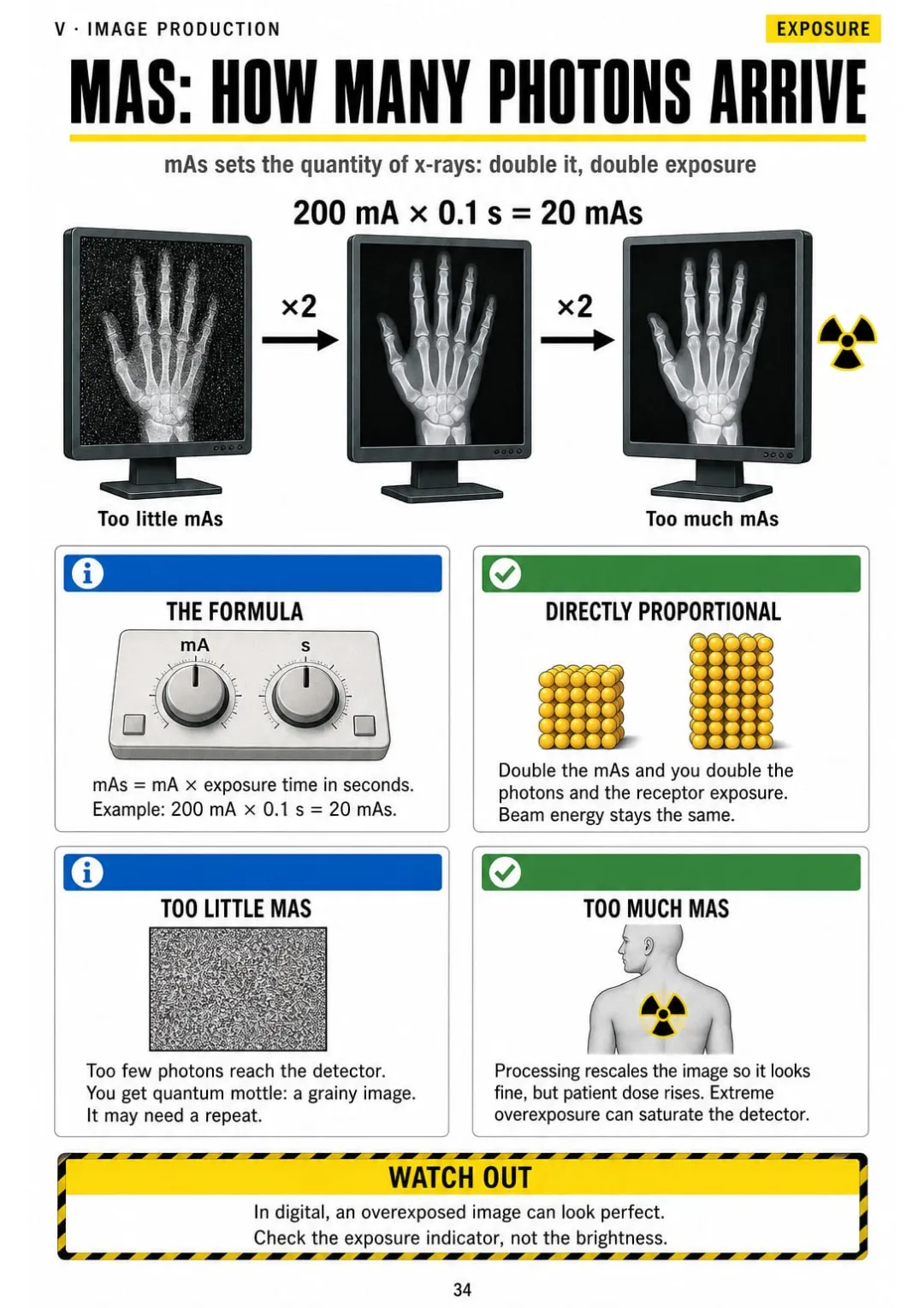
mAs, kVp and the 15% rule, SID, resolution, distortion, scatter, grids and cutoff, CR vs DR, the exposure indicator, artifacts and AEC: what to change when an image comes out wrong.
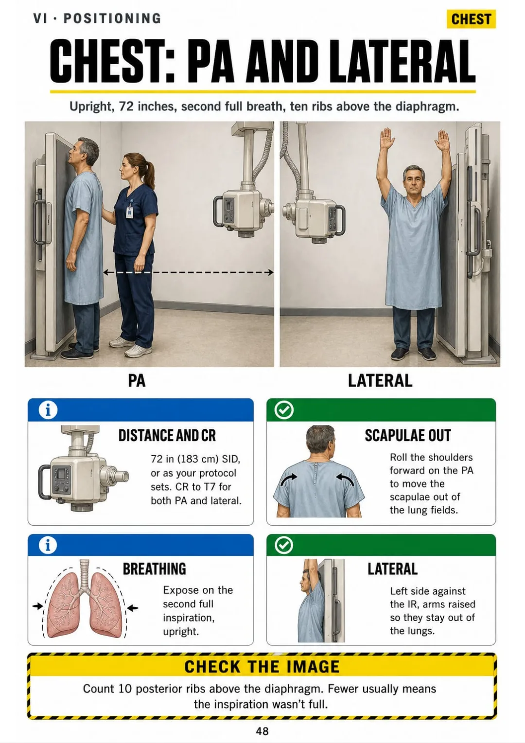
Projection vs position, surface landmarks and every routine body part from chest and abdomen to spine and skull, plus trauma, mobile, pediatric and contrast studies: the central ray, the angle and the landmark drawn on the body.
All the calculations on one page, the numbers to know cold, the most frequent mistakes side by side and a week-before checklist.
At the end of the book: All the calculations on one page, the numbers to know cold, the most frequent mistakes side by side and a week-before checklist.

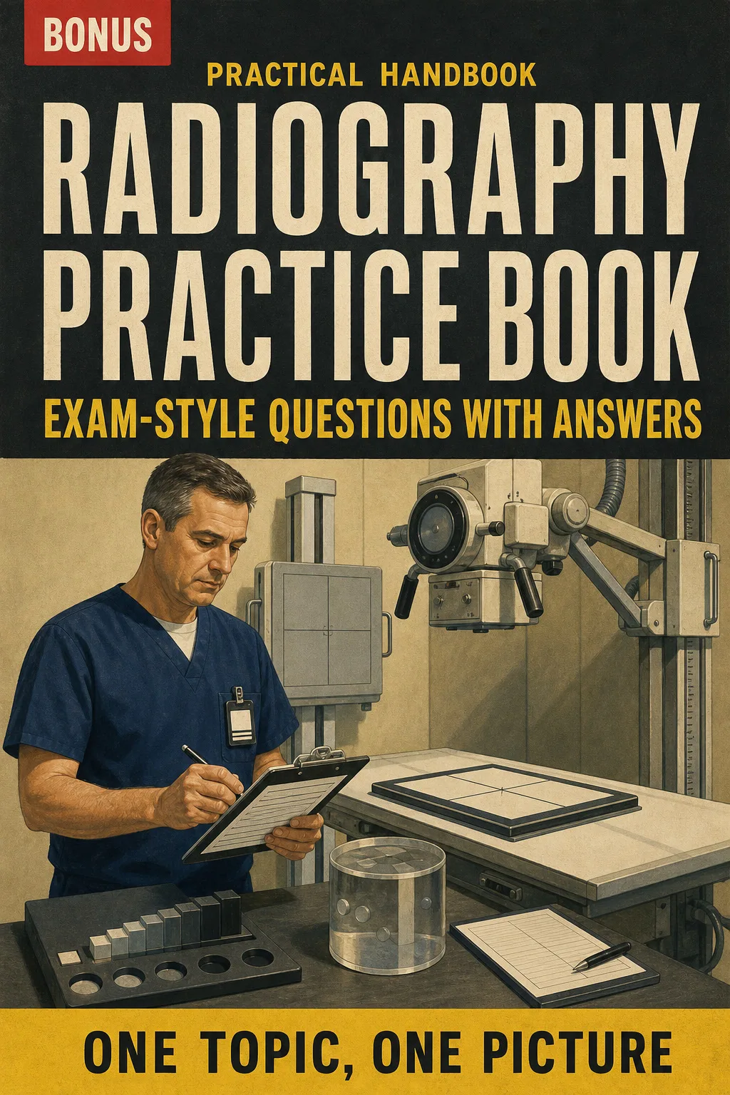
Test yourself on every content area and see why each answer is right, with the page to go back to.

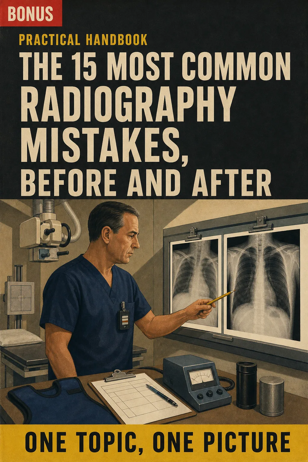
Spot the errors that trip students up (distance, angles, exposure, shielding) and see the fix drawn side by side.
📄 This is a digital PDF book sent to your email: nothing is printed or shipped.
PDF by email: We email you Radiography Exam Illustrated as a PDF, together with the two free books. It's a single payment: no installments, no extra charges.
Every lecture topic has a page you can review the same night, with the drawing that makes the physics click.
You review the whole content outline in order, one topic per page, and solve every calculation in the same 4 steps.
Print the positioning pages and keep them at hand: the central ray, the angle and the landmark drawn on the body.
📄 This is a digital PDF book sent to your email: nothing is printed or shipped.
Try Radiography Exam Illustrated for 30 days, bonus books included.
We refund everything if the file won't open, if the book isn't what you see here, if you were charged twice or if the email doesn't arrive. Just email us.
You pay once. No subscriptions.
Yes. It doesn't replace them: your app tests you, the textbook explains in paragraphs, and this book puts each topic on one drawn page with the numbers, angles and steps in a box. It isn't a beginner's book: it's built to review the whole content outline of the US radiography certification exam in order, in the last weeks before your test date.
It follows the ARRT Radiography Examination Content Specifications in effect since January 2022, organized in the same 4 areas: Patient Care, Safety, Image Production and Procedures. Dose limits and equipment limits are checked against NCRP Report No. 116, NRC 10 CFR Part 20 and FDA 21 CFR Part 1020. Before your exam, check the ARRT website for the specifications that apply to your test date.
No. It's independent study material, not affiliated with or endorsed by ARRT, NCRP, NRC or FDA.
No. It's a study and review tool. It doesn't replace your program, your clinical training or your hospital's protocols, and it doesn't give you a certificate or CE credits.
No. They're written for this book, following the exam content areas, and they aren't official questions from any exam.
The book and the 2 bonus books arrive as PDFs by email, at the address you enter at checkout.
No. It's a one-time payment.
Yes. It's a PDF you can print, all of it or just the pages you want to keep at hand in clinicals.
You have 30 days. We refund everything if the file won't open, if the book isn't what you see on this page, if you were charged twice or if the email doesn't arrive: just email us.
No more rereading without anything sticking.
The central ray and the angle: you see them drawn, and that's it.
You walk into the exam knowing where everything is.
Drawings make studying faster.
The book is right below.
One-time payment: US$ 37 · Guarantee: 30 days · PDF by email
YES, I WANT RADIOGRAPHY EXAM ILLUSTRATED FOR US$ 37📄 This is a digital PDF book sent to your email: nothing is printed or shipped.